Featured

High school teachers learn about electrical circuits using water analogies
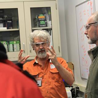
Even a Saturday snow storm wasn’t enough to keep science teachers away from a teacher workshop at CHESS this past March (even the ones that drove all the way from Virginia!).
Lateral or vertical — that is the question!
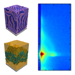
In a Forum Article, which was recently published online in the journal “ACS Applied Materials & Interfaces” [1], Anatoly V. Berezkin and coworkers attempted to figure out what happens when you prepare thin films from two homologous diblock copolymers differing only in overall length.
March 2017 workshop on CHESS-U as a pulsed x-ray source
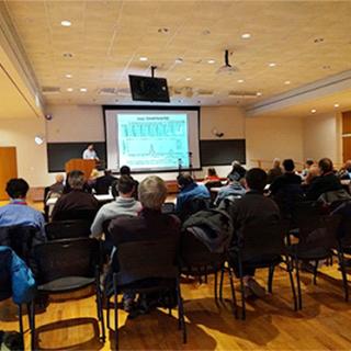
Our users may be aware that during the summer of 2016, CHESS hosted six workshops to help outline a science case for an upgraded single-beam, high-energy source that will result from the current CHESS-U upgrade project.
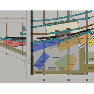
The state of CHESS-U beamlines
The beamline upgrade portion of the CHESS-U project is moving ahead at full steam. Scientific needs have been identified. A suitable layout for the experimental floor has been devised.
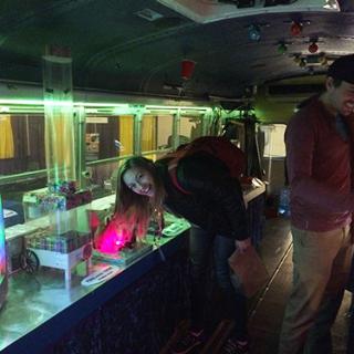
Physics Bus magnetism attracts students to science at New Orleans APS meeting in March
CHESS enjoyed a strong presence at the March APS meeting in New Orleans last month.
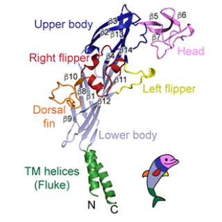
The Big Pore Theory could cure chronic pain
Cornell University researchers have produced for the first time an image of P2X7, a receptor associated with chronic pain.
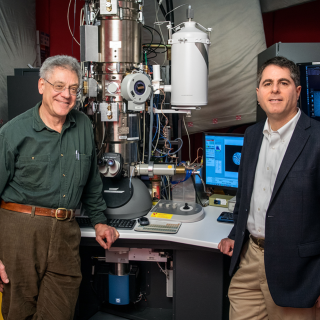
New electron microscope sees more than an image
The electron microscope, a powerful tool for science, just became even more powerful, with an improvement developed by Cornell physicists. Their electron microscope pixel array detector (EMPAD) yields not just an image, but a wealth of information about the electrons that create the image and, from that, more about the structure of the sample.
Researchers look for genetic clues to help grapes survive cold
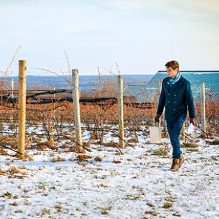
Months before northern vineyards burst into their lush summer peak, the delicate grape buds holding the nascent fruit in its tiny core must first withstand the freezing onslaught of winter.