Home
Home
  • Status
  • Science
    • Art and Archaeology
    • Biology
    • Chemistry
    • Energy
    • Engineering
    • Materials
    • X-Ray Technology
    • User Stories
    • Science Highlights
    • Publications
  • Users
    • 2025 CHESS User Meeting
    • Workshops at CHESS
    • Beamline Directory
    • X-Ray Run Schedule
    • What's the process? - Prospective User Guide
    • User Guide
    • User Agreement
    • BeamPASS
    • CHESS Deadlines
    • CHESS Status Page
    • Safety
      • In-Person User Orientation and Safety Training
    • Technical Resources
      • Affiliated Resources
      • Calculators
      • Computing
      • Detectors
      • Video Backgrounds
    • Acknowledgments
    • Travel and Lodging
    • Shipping
  • Facilities
    • Becoming a Partner
    • CHEXS
    • HMF Beamline
    • MSN-C
    • MacCHESS
    • XLEAP
      • People of XLEAP
      • XLEAP Overview
      • Proposed Capabilities
      • Stay in touch
  • Public
    • Events
    • Tours
    • Student Opportunities
    • Lending Library
    • 3D and Virtual Tours
  • Industry
  • About
    • Staff Directory
    • Advisory Bodies
    • What we do
    • Job Openings
    • News
      • CHESS eNewsletter
      • Media Resources
      • News Archive
    • Publications
      • Publications 2025
      • Publications 2024
      • Publications 2023
      • Publications 2022
      • Publications 2021
      • Publications 2020
      • Publications 2019
      • Publications 2018
      • Publications 2017
      • Publications 2016
      • Publications 2015
      • Publications 2014
      • Publications 2013
      • Publications 2012
      • Publications 2011
      • Publications 2010
      • Publications 2009
      • Publications 2005
    • Beyond the Lab
    • History
Science Highlights at CHESS

Science Highlights

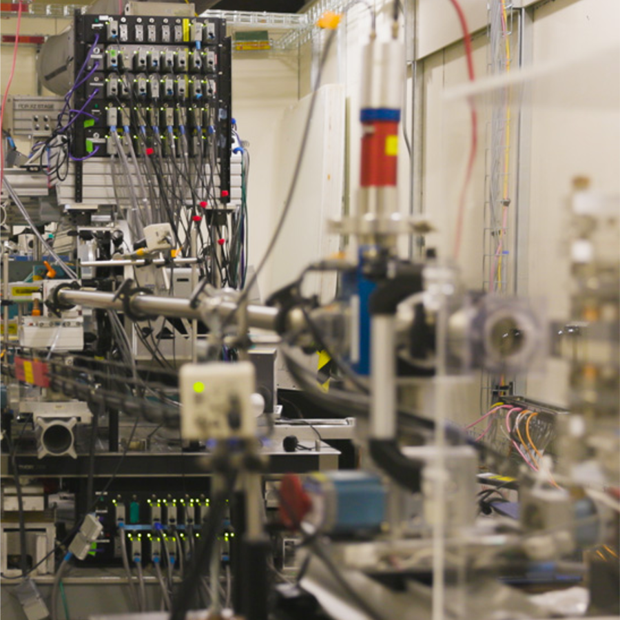
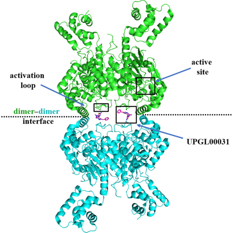
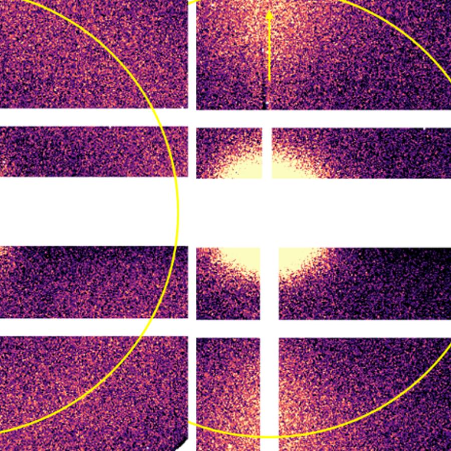
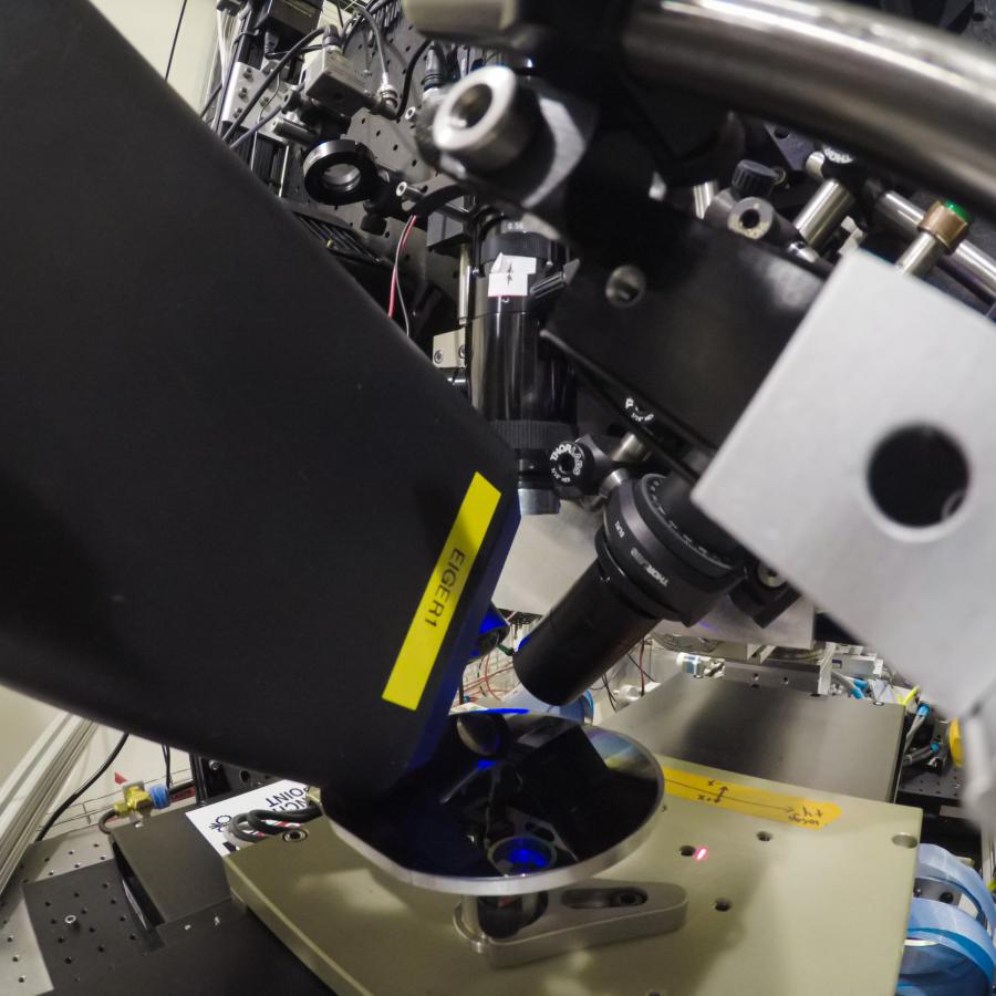
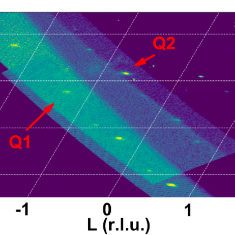
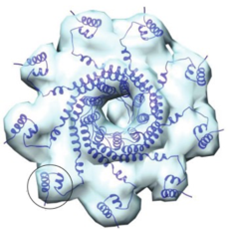
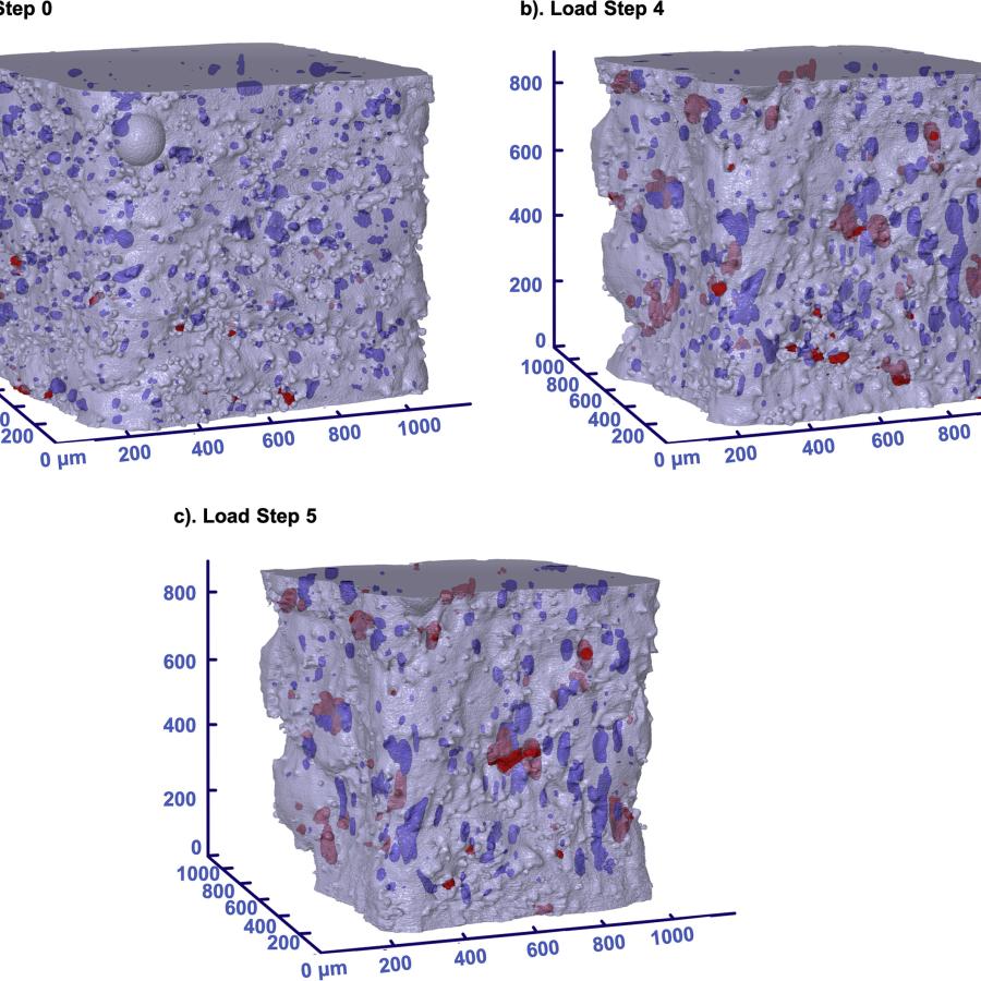
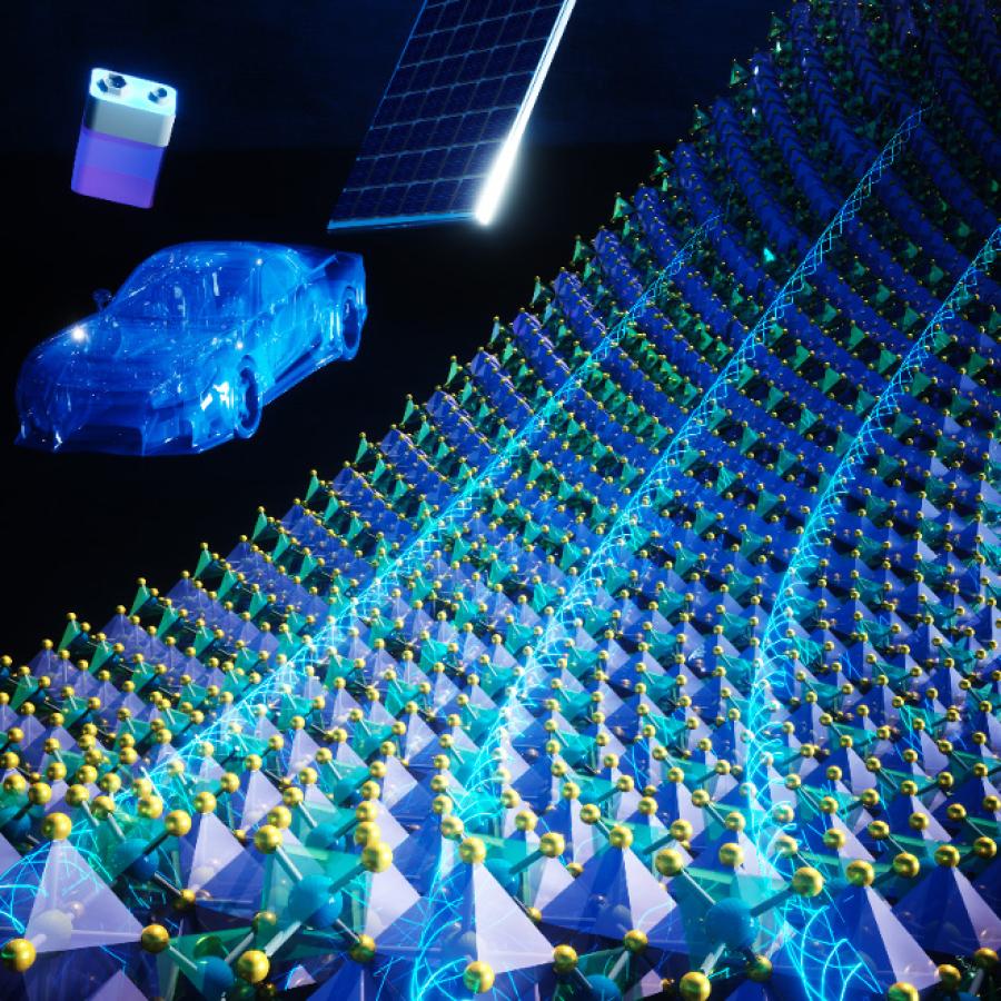
At CHESS, X-rays are used for examining the structure and function of our material world, both living and not, with an eye towards designing new materials, solving technological problems, and even curing diseases.

Energy vs Normalized Absorption

Redox Mechanisms and Migration Tendencies in Earth-Abundant Cathodes

HiP dual E loop

Pushed to extremes: distinct effects of high temperature versus pressure on the structure of STEP

Triangular lattice

Frustrated charge order and cooperative distortions in ScV6Sn6

graph of particle trajectories

X-ray imaging of powder particles driven from a surface by a nearby electrostatic discharge

HEDM image, larger image and description below.

Intragranular micromechanical fields at triple junctions

image description in caption

Residual Stress Model Validation of Cold Hole Expansion in an Aerospace Aluminum Alloy

""

New Methodology to Collect X-ray Emission Spectroscopic Data for Platinum and Other Heavy Metals

image showing an artistic rendering of diffraction

Machine learning enhances X-ray imaging of nanotextures

Portrait of Catherine Royer

CHESS Research Uses Pressure To Understand RNA Dynamics

Image description in caption.

The Functional Materials Beamline in SRN

Diagram of diffuse scattering variations

Picking up good vibrations – of proteins – at CHESS

Image of multicolored HEDM characterization of grain structures, description in caption.

Grain scale residual stress after quasi-static and high strain rate loading in SS316L

image of rainbow-colored shapes

Using real-time data analysis to conduct next-generation synchrotron fatigue studies

Image of a high pressure sample setup.

Inline small-angle X-ray scattering-coupled chromatography under extreme hydrostatic pressure

abstract image of blue and pink graphs

The influence of Alloying on slip intermittency and the implications for dwell fatigue in titanium

image of enzyme molecules

Crystal structure of a type III Rubisco in complex with its product 3-phosphoglycerate

Tree inference diagram for RNR sequence

Protein family shows how life adapted to oxygen

pressure picture

Nanocrystals with Metastable High-Pressure Phases Under Ambient Conditions

the sccu undulator

Super Cornell Compact Undulator (sCCU) Compact Variable-Gap Undulator with Hydraulic-Assist Driver and Enhanced Magnetic Field

imag description in caption

Mapping Liquid Formulations with the Autonomous Formulation Laboratory at CHESS-FMB

image of a portion of an intensity chart

Intermediate Valence State in YbB4 Revealed by Resonant X-ray Emission Spectroscopy

a cropped portion of diffraction images

Dynamics of Recrystallized Grains in a Hot-Compressed Mg-Zn-Ca Alloy

image of a person reflected in a detector

Characterization of 128 x 128 MM-PAD-2.1 ASIC: a Fast Framing Hard X-ray Detector with High Dynamic Range

image of a graph showing two descending lines

Unconventional Hysteretic Transition in a Charge Density Wave

image of the PIPOXS beamline

Nonprecious transition metal nitrides as efficient oxygen reduction electrocatalysts for alkaline fuel cells

abstract green and blue image

Analysis of a three-dimensional slip field in a hexagonal Ti alloy from in-situ high-energy X-ray diffraction microscopy data

Image description in caption

How two cancer drugs can look the same but behave differently - revealed by serial room temperature crystallography

abstract image of x-ray scattering

Measuring complex fluids under extreme flow conditions

Green tea

BioSAXS helps to explain the anti-cancer activity of green tea

Image of researchers at the Functional Materials Beamline

In situ SAXS/WAXS mapping of thermoplastic crystallization during 3D printing

na

Assessment of DFT methods for geometry optimization of platinum-containing complexes

image of scattering chamber schematic, description in caption

Testing for the Continuous Spectrum of X-Rays Predicted to Accompany the Photoejection of an Atomic Inner Shell Electron 

image of lattice strains, description in caption

Phase-specific deformation in additively manufactured Ni–CrC composites

image of Iridium trimer compound Ba4Ir3O10, description in caption

Structural and magnetic transitions in the planar antiferromagnet Ba4Ir3O10

image of a red, white and blue beta-lactoglobulin 3D structure

Progress in high-pressure crystallography at ID7B2 furthers the mission of HP-Bio

an image of indexed grains, description in caption

Grain-scale deformation of a high entropy alloy using synchrotron high energy diffraction microscopy

Image shows a figure of an electrochemical cell.

In situ spectroscopy as a probe of electrocatalyst performance

Image shows 3D and 2D presentations of date. Description in caption

Unconventional chiral charge order in kagome superconductor KV3Sb5

Kelly Nygren of CHESS loads a sample into RAMS

In-Hutch Commissioning of Rotation and Axial Motion System IV (RAMS IV) Load Frame

protein

Protein unfolded states populated at high and ambient pressure are similarly compact

Sydor detector at CHESS beamline

Next-generation detectors ready for experiments at CHESS

laser annealing setup with detector

Autonomous materials development using in situ laser annealing and scan-probe, grazing incident x-ray microdiffraction.

Showing the subtle superstructure at low temperatures of Ta4Pd3Te16

Incommensurate charge order in a low-dimensional superconductor

bacteriophage as described in caption

A virus recognizes the starting point on the DNA to be packaged inside its protein shell

image shows pores evolving, description in caption.

Investigation of porosity, texture, and deformation behavior using high energy X-rays during in-situ tensile loading in additively manufactured 316L stainless steel

High-stress steel microstructure

Quantifying Through-Thickness Residual Stresses from Forming of Wrought Steel Armor Plate

Three-dimensional renderings of the sample volumes grown from the centroids measured using ff-HEDM.

Grain-resolved temperature-dependent anisotropy in hexagonal Ti-7Al revealed by synchrotron X-ray diffraction

graph of plasmon intensity and Fe2+ molar percentage

Fe Cations Control the Plasmon Evolution in CuFeS2 Nanocrystals

antifluorite structure

Approaching the ideal limit for spin-orbit-coupled quantum moments in iridium halides

HPSAXS Cell at CHESS

High-pressure small-angle X-ray scattering cell for biological solutions and soft materials

Gabrielle Illava manipulating samples within the anaerobic chamber

BioSAXS facility at CHEXS develops stringent anoxic sample environment

clog development sequence

Mechanics of nozzle clogging during direct ink writing of fiber-reinforced composites

grains in the HEDM

X-rays quantify stress gradients at twin boundaries, which can drive fatigue failure in nickel-based superalloys.

Ivan and Alexander Temnykh

Developing the Next Generation of CHESS Compact Undulators

SAXS intensity map reveals structure of 3D-printed part

Revealing Filler Morphology in 3D-Printed Thermoset Nanocomposites by Scanning Microbeam X-ray Scattering

HEDM initial orientation

In-situ high energy X-ray diffraction probes elastic response of metastable engineered alloys

Beam through Steel

Validation of Welding Model for Digital Twin of Thick-Plate Joint: Mapping Residual Strains in HSLA Steel using High Energy X-rays

updated small-polaron hopping model

Breakdown of the Small-Polaron Hopping Model in Higher-Order Spinels

unmodulated structure

X-rays uncover “hidden” quantum states

schematic of storage ring

Minimizing Deviations: Improving Beam Stability at CHESS

View into the diamond anvil cell

Biology Under Pressure: Beta-lactoglobulin survives under pressures as high as 9000 bar

nickel-based superalloy sample

Unsupervised Learning of Dislocation Motion

Schematics of bunch patterns

Development of new bunch pattern at CHESS for dynamics studies

EXAFS Data

2020 PREM X-ray Absorption Spectroscopy and EXAFS Workshop

RNA

RNA's magic begins with base-specific ion interactions

cryo-TEM images

Crystallization-Driven Self-Assembly of Coil−Comb-Shaped Polypeptoid Block Copolymers

Schematic of the experimental setup for the collection of both small angle scattering (SAXS) and wide angle scattering (WAXS) images from nanocrystal assemblies. The top panel illustrates the different length scales determining the characteristics of the individual nanocrystals and the supercrystals

Fabricating, Characterizing and Understanding Nanocrystal Superstructures

Small-angle X-ray scattering (SAXS) setup and analysis for a nanocomposite polymer.

Preparation and Characterization of Mesoscale Single Crystals

Cover of Nature

Spontaneous Gyrotropic Electronic Order in 1?-TiSe₂

A highly detailed three-dimensional map of diffuse scattering

Diffuse X-ray Scattering from Correlated Motions in a Protein Crystal

Schematic time dependence of caging strength in self-suspended hairy nanoparticles upon annealing.

Thermally Activated Equilibration of Self-Suspended Hairy Nanoparticles

Otoliths and corresponding chemical profiles of two specimens performing regional natal homing in the upper Madeira River

Unmasking continental natal homing in goliath catfish from the upper Amazon

Experimental X-ray scattering curves for RNA duplexes

Twisting the Helix: Salt Dependence of Conformations of RNA Duplexes

TEM image of a typical PbSe nanocrystal

Setting Carriers Free: Healing Faulty Interfaces Promotes Delocalization and Transport in Nanocrystal Solids

Growth pattern of caprine horn.  Left:  Diagram of a horn for a 5-year old goat indicating the layer of horn grown at each year of age. Right: Example of a horn cross section, indicating the layer of horn grown at each year of age.

Lead uptake and distribution in horns from goats

Evolution of diffracted intensity from basal lattice planes

To Twin or Not to Twin: Micromechanical Response in Magnesium probed with High Energy X-Rays

Schematic of the in situ XAS electrochemical cell.

Synergistic Co−Mn Oxide Catalyst for Oxygen Reduction Reactions

Left: The cellulose fibers are naturally aligned along the growth direction. Cellulose exhibits a hierarchical alignment, in which the nanofibres consist of aligned cellulose molecular chains. Right: Schematic of the ionic device after infiltrating electrolyte into the nanofluidic cellulosic membrane. Under a thermal bias, the surface charged nanofibres regulate ionic movement via nanofluidic effects (that is, the overall effect created by a high degree of confinement and the charged walls of the cellulose

Heat Harvesting with Cellulosic Membranes

Schematic side view (top) and photographic top view (bottom)of the transmission mirror test set up. A comparison of the reflected intensity of the tungsten Lα, Lβ, and Lγ lines provided by a microfoucs tube source were used to quantify the rejection ratio of the XTM for different angles of incidence.

Potential high-yield fabrication of x-ray transmission mirror optics

dual array valence emission spectrometer

Hopping Made Easy: Controlling Electronic Transport in CoₓMn₃₋ₓO₄ Nanoparticles

Profile of X-ray beam reflected from a CVD

Diamonds are for focusing and monochromatizing: Chemically vapor deposited diamond crystal as medium resolution X-ray monochromator

spin stripes

Less is more: Disorder raises the critical temperature of a cuprate superconductor

Small-angle X-ray scattering

Conformational Gymnastics Necessary for Ribonucleotide Reductase Activity

Serial crystallography

Room temperature serial oscillation crystallography

Controlling Lithium Dendrite Growth: An Operando X-ray Study

Science HighlightYbRu2Ge2

Interplay of electronic and structural degrees of freedom in YbRu2Ge2

Science Highlight

Probing the Micromechanical Response of Crystalline Phases in Alternate Cementitious Materials using 3-D X-ray Techniques

Science Highlight at CHESS

Relation of local order to material properties in ferroelectrics

insitu observations fast

In-Situ Observations of Performance Evolution in Shape Memory Alloys

Footer menu

  • Newsletter
  • CLASSE
  • Contact
  • Staff
  • Feedback
  • Web Accessibility Help
The Cornell High Energy Synchrotron Source (CHESS) is operated and managed by Cornell University.
CHESS/Wilson Lab 161 Synchrotron Drive Ithaca, NY 14853
© 2025 Cornell High Energy Synchrotron Source